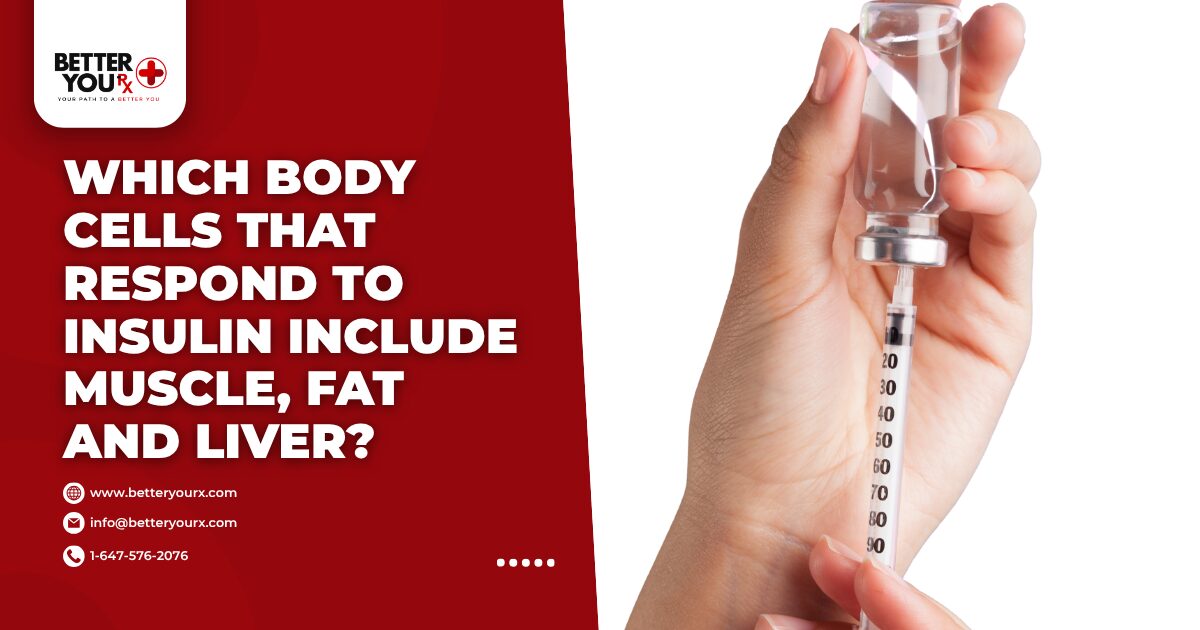
Body cells that respond to insulin include muscle cells, fat cells, and liver cells as the primary targets for glucose uptake and metabolism. These insulin-sensitive cells contain special receptors that bind with insulin molecules, triggering processes that allow glucose to enter from the bloodstream. Brain cells, kidney cells, and blood vessel cells also respond to insulin signals, though their glucose uptake mechanisms differ. Each cell type plays a unique role in maintaining blood sugar balance throughout the body. Insulin resistance occurs when these normally responsive cells stop reacting properly to insulin signals, leading to elevated blood glucose levels and potential diabetes development.
Primary Body Cells That Respond to Insulin Include Muscle Tissue
Muscle cells represent the largest insulin-responsive tissue in the human body, accounting for approximately 80% of glucose disposal after meals. These cells contain abundant insulin receptors on their surface membranes that activate when insulin binds to them.
Skeletal Muscle Function
Skeletal muscles use glucose for immediate energy during physical activity and store excess glucose as glycogen for future use. When insulin attaches to muscle cell receptors, it triggers a cascade of cellular events.
- GLUT4 transporters move from inside the cell to the surface membrane
- Glucose channels open to allow sugar molecules inside
- Enzymes activate to convert glucose into glycogen
- Protein synthesis increases for muscle repair and growth
- Fat oxidation adjusts based on available glucose
- Amino acid uptake enhances for tissue building
Regular physical activity improves muscle insulin sensitivity, meaning cells respond more efficiently to insulin signals. This enhanced responsiveness explains why exercise helps control blood sugar levels in people with diabetes.
Cardiac Muscle Response
Heart muscle cells continuously require energy and respond readily to insulin for glucose uptake. The cardiac tissue maintains its own glycogen stores while primarily using fatty acids for energy between meals.
Insulin affects heart muscle by promoting glucose utilization during the fed state, protecting against lipid accumulation, and supporting proper cardiac contractility. Heart cells express high levels of insulin receptors, making them particularly sensitive to insulin availability and function.
Smooth Muscle Involvement
Smooth muscle cells lining blood vessels and internal organs also respond to insulin, though less dramatically than skeletal muscle. These cells help regulate blood pressure and organ function.
- Vascular smooth muscle relaxes in response to insulin
- Digestive tract muscles coordinate better with insulin present
- Bladder and reproductive organ muscles maintain proper tone
- Blood flow increases to insulin-sensitive tissues
- Nutrient delivery improves throughout the body
How Fat Cells React to Insulin Signals

Adipose tissue serves as the body’s primary energy storage system, with fat cells responding to insulin by taking up glucose and converting it into triglycerides for long-term storage.
White Adipose Tissue
White fat cells specialize in energy storage and release, responding to insulin by suppressing lipolysis and promoting lipogenesis. These cells can expand significantly to accommodate excess energy storage.
Insulin triggers several processes in white fat cells. First, it activates lipoprotein lipase on the cell surface, which breaks down circulating triglycerides for uptake. Second, it stimulates glucose uptake through GLUT4 transporters similar to muscle cells. Third, it promotes the conversion of glucose into fatty acids through de novo lipogenesis.
| Fat Cell Response | Effect on Metabolism |
| Increased glucose uptake | Lower blood sugar |
| Enhanced fat storage | Reduced circulating lipids |
| Decreased fat breakdown | Stable energy reserves |
| Adipokine secretion | Whole-body insulin sensitivity |
| Cell size regulation | Metabolic health markers |
Brown Adipose Tissue
Brown fat cells generate heat through thermogenesis and respond to insulin differently than white fat cells. These specialized cells contain numerous mitochondria that burn glucose and fatty acids to produce warmth rather than storing energy.
- Higher metabolic rate than white fat
- Increased glucose consumption for heat production
- Enhanced mitochondrial function with insulin signaling
- Greater insulin receptor density
- Direct contribution to glucose homeostasis
- Protection against metabolic dysfunction
Beige Adipocytes
Beige fat cells represent an intermediate type between white and brown adipose tissue. These cells can switch between energy storage and heat production based on metabolic needs and insulin signaling.
Cold exposure and exercise can convert white fat cells into beige adipocytes, improving overall insulin sensitivity. This browning process enhances the body’s ability to regulate blood sugar and burn excess calories.
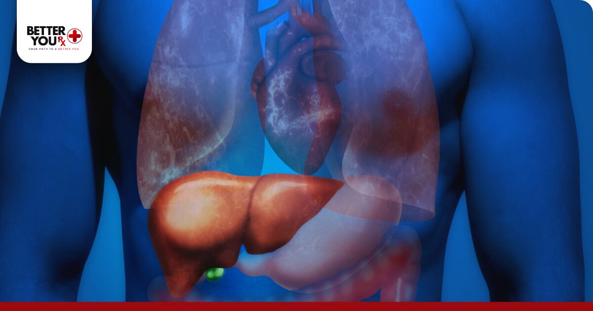
Liver Cells and Glucose Metabolism
The liver acts as the body’s glucose buffer, with hepatocytes responding to insulin by shifting from glucose production to glucose storage and lipid synthesis.
Hepatocyte Insulin Response
Liver cells contain abundant insulin receptors that regulate multiple metabolic pathways simultaneously. When insulin levels rise after eating, hepatocytes immediately respond by changing their metabolic priorities.
Insulin suppresses gluconeogenesis, the process of creating new glucose from amino acids and other substrates. This prevents the liver from adding more glucose to already elevated post-meal blood sugar levels. Simultaneously, insulin activates glycogen synthase, directing incoming glucose toward glycogen storage.
- Glucose uptake increases through GLUT2 transporters
- Glycogen synthesis accelerates for energy storage
- Fat production begins from excess carbohydrates
- Protein synthesis enhances for cellular maintenance
- Inflammatory markers decrease with proper signaling
- Detoxification processes improve with adequate insulin
Glycogen Storage Regulation
The liver stores approximately 100-120 grams of glycogen, providing readily available glucose between meals. Insulin precisely controls this storage process through multiple enzymatic pathways.
During feeding, insulin promotes glycogen synthesis by activating glycogen synthase and inhibiting glycogen phosphorylase. This dual regulation ensures efficient glucose storage when blood sugar levels are elevated. The liver can store enough glycogen to maintain blood glucose for 12-24 hours of fasting.
Get reliable diabetes management with Humalog Junior KwikPen from Better You Rx, ensuring precise insulin delivery for optimal cellular response.
Lipid Metabolism Control
Beyond glucose regulation, liver cells respond to insulin by altering fat metabolism. Insulin promotes de novo lipogenesis, converting excess glucose into fatty acids for export to adipose tissue.
| Liver Function | Insulin Effect |
| Glucose production | Strongly suppressed |
| Glycogen synthesis | Markedly increased |
| Fat synthesis | Stimulated |
| Cholesterol production | Enhanced |
| Protein synthesis | Promoted |
| VLDL secretion | Regulated |
This coordinated response prevents excessive glucose accumulation while supporting whole-body energy balance.
Which Body Cells That Respond to Insulin Include Kidney and Brain Tissues?
While muscle, fat, and liver cells are primary insulin targets, other tissues throughout the body also respond to insulin signals in important ways for maintaining metabolic health.
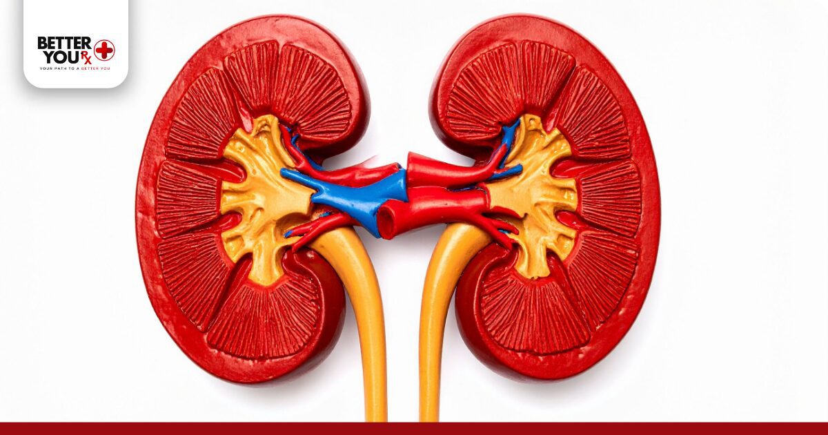
Kidney Cell Response
Kidney cells express insulin receptors and play crucial roles in glucose homeostasis beyond simple filtration. The kidneys both reabsorb filtered glucose and produce new glucose through gluconeogenesis.
Proximal tubule cells respond to insulin by increasing glucose reabsorption through sodium-glucose cotransporters. This prevents glucose loss in urine when blood sugar levels are normal. Insulin also affects kidney gluconeogenesis, though less dramatically than in the liver.
- Enhanced sodium reabsorption affecting blood pressure
- Increased glucose threshold for urinary excretion
- Improved protein handling and reduced loss
- Better electrolyte balance maintenance
- Reduced inflammation in kidney tissue
- Protection against diabetic kidney disease progression
Brain Tissue Considerations
Brain cells have a complex relationship with insulin, as neurons can take up glucose independently of insulin through GLUT1 and GLUT3 transporters. However, specific brain regions do respond to insulin signals.
The hypothalamus contains insulin-sensitive neurons that regulate appetite, energy expenditure, and glucose metabolism throughout the body. Insulin signaling in the brain affects memory formation, cognitive function, and mood regulation. The hippocampus particularly benefits from proper insulin signaling for memory consolidation.
Astrocytes and other supporting brain cells also respond to insulin, helping maintain the blood-brain barrier and supporting neuronal function. This insulin sensitivity in brain support cells contributes to overall cognitive health and protection against neurodegenerative conditions.
Endothelial Cells and Vascular Response
Blood vessel lining cells respond to insulin in ways that affect circulation, blood pressure, and nutrient delivery to all tissues.
Vascular Endothelium Function
Endothelial cells line all blood vessels and respond to insulin by producing nitric oxide, a molecule that causes blood vessel dilation. This vasodilation improves blood flow to insulin-sensitive tissues, enhancing glucose uptake and utilization.
Healthy insulin signaling in endothelial cells maintains proper vascular function through multiple mechanisms. Nitric oxide production increases, reducing blood pressure and improving tissue perfusion. Anti-inflammatory pathways activate, protecting against atherosclerosis development.
- Enhanced blood flow to skeletal muscle
- Improved nutrient delivery to all tissues
- Reduced platelet aggregation and clotting risk
- Decreased vascular inflammation markers
- Better oxygen distribution throughout body
- Protection against endothelial dysfunction
Microvascular Responses
Small blood vessels and capillaries show particular sensitivity to insulin, with effects on tissue nutrition and waste removal. Insulin increases capillary recruitment in muscle tissue, expanding the surface area available for nutrient exchange.
This microvascular response to insulin occurs rapidly, often preceding measurable glucose uptake in muscle tissue. Impaired microvascular insulin signaling contributes to insulin resistance development and diabetes complications affecting eyes, kidneys, and nerves.
Pancreatic Cell Types and Insulin Interaction
The pancreas contains multiple cell types that both produce and respond to insulin, creating complex feedback loops for glucose regulation.
Beta Cell Function
Pancreatic beta cells produce and secrete insulin while also responding to their own insulin signals through autocrine mechanisms. These cells contain insulin receptors that provide feedback about insulin production needs.
Beta cells sense blood glucose through GLUT2 transporters and glucokinase enzymes. When glucose enters beta cells, it triggers metabolic changes leading to insulin secretion. The cells then respond to secreted insulin by modulating their own function and survival.
Consider Lantus SoloStar for consistent basal insulin coverage, supporting natural cellular insulin response patterns throughout the day.
Alpha Cell Regulation
Alpha cells produce glucagon, insulin’s counter-regulatory hormone, and express insulin receptors that suppress glucagon secretion when insulin levels rise. This paracrine signaling between beta and alpha cells fine-tunes glucose homeostasis.
- Insulin directly inhibits glucagon release
- Prevents inappropriate glucose production
- Maintains balanced hormone secretion
- Coordinates islet cell function
- Protects against hypoglycemia
- Supports metabolic flexibility
Delta and PP Cells
Delta cells secrete somatostatin, which modulates both insulin and glucagon secretion. These cells respond to local insulin concentrations by adjusting somatostatin output, creating additional regulatory control.
Pancreatic polypeptide cells (PP cells) also express insulin receptors and contribute to appetite regulation and digestive function. The coordinated response of all pancreatic cell types ensures precise glucose control under varying metabolic conditions.
Immune Cells and Inflammatory Response
Immune system cells express insulin receptors and respond to insulin signals in ways that affect inflammation and metabolic health.
Macrophage Insulin Sensitivity
Macrophages in adipose tissue and other organs respond to insulin by shifting from pro-inflammatory to anti-inflammatory states. Proper insulin signaling in macrophages reduces cytokine production and improves tissue insulin sensitivity.
Insulin affects macrophage polarization, promoting the M2 anti-inflammatory phenotype over the M1 pro-inflammatory state. This shift reduces chronic inflammation associated with obesity and metabolic syndrome.
| Immune Cell Type | Insulin Response |
| Macrophages | Reduced inflammation |
| T cells | Improved regulation |
| B cells | Enhanced antibody production |
| Neutrophils | Decreased activation |
| Dendritic cells | Better antigen presentation |
| Natural killer cells | Modulated activity |
Lymphocyte Function
T cells and B cells express insulin receptors that influence their activation, proliferation, and function. Insulin signaling supports appropriate immune responses while preventing excessive inflammation.
Insulin affects T cell metabolism, shifting from glycolysis to oxidative phosphorylation for more efficient energy production. This metabolic flexibility allows proper immune function without excessive inflammatory responses that could impair insulin sensitivity in other tissues.
Bone Cells and Metabolic Regulation
Skeletal system cells respond to insulin in ways that affect both bone health and whole-body metabolism.
Osteoblast Activity
Bone-forming osteoblasts express insulin receptors and respond by increasing bone formation and secreting osteocalcin, a hormone that enhances insulin sensitivity in other tissues. This creates a feedback loop between bone and energy metabolism.
Insulin signaling in osteoblasts promotes collagen synthesis and mineralization while supporting bone density maintenance. The osteocalcin released by these cells travels to muscle and fat tissue, improving their insulin responsiveness.
- Enhanced bone formation and strength
- Increased osteocalcin production
- Improved whole-body insulin sensitivity
- Better calcium homeostasis
- Reduced fracture risk
- Support for metabolic health
Osteoclast Regulation
Bone-resorbing osteoclasts also respond to insulin, though primarily through indirect mechanisms. Insulin affects osteoclast differentiation and activity through its effects on osteoblasts and immune cells.
Proper insulin signaling maintains balanced bone remodeling, preventing excessive bone loss while allowing necessary turnover for skeletal health. This balance becomes particularly important with aging and metabolic dysfunction.
Reproductive System Cells
Reproductive organs contain insulin-responsive cells that link metabolic health with fertility and hormone production.
Ovarian Cell Response
Ovarian cells, including granulosa and theca cells, express insulin receptors that influence hormone production and egg development. Insulin works synergistically with reproductive hormones to regulate ovarian function.
Excessive insulin signaling in ovaries can disrupt normal hormone production, contributing to conditions like polycystic ovary syndrome. Balanced insulin action supports regular ovulation and appropriate sex hormone levels.
- Follicle development regulation
- Estrogen and progesterone production
- Egg maturation support
- Menstrual cycle coordination
- Fertility optimization
- Hormone balance maintenance
Testicular Function
Leydig cells in testes respond to insulin by modulating testosterone production. Proper insulin signaling supports normal testosterone levels and sperm production.
Sertoli cells, which nurture developing sperm, also express insulin receptors. Insulin affects their function in supporting spermatogenesis and maintaining the blood-testis barrier. Metabolic health through proper insulin signaling directly impacts male fertility and hormone balance.
Achieve better glucose control with NovoRapid FlexPens, helping your body’s cells respond optimally to insulin signals.
Intestinal Cells and Nutrient Absorption
Digestive system cells respond to insulin in ways that coordinate nutrient absorption with metabolic needs.
Enterocyte Response
Intestinal absorptive cells express insulin receptors that influence nutrient uptake and processing. Insulin affects the expression of nutrient transporters and digestive enzymes.
These cells increase glucose and amino acid absorption in response to insulin while also secreting incretin hormones that enhance insulin secretion from pancreatic beta cells. This gut-pancreas axis ensures coordinated responses to food intake.
Enteroendocrine Cells
Specialized intestinal cells that produce hormones respond to both nutrients and insulin. These cells secrete GLP-1, GIP, and other hormones that regulate insulin secretion and appetite.
- Enhanced incretin hormone production
- Improved glucose-stimulated insulin release
- Better appetite regulation
- Coordinated digestive function
- Reduced inflammation in gut tissue
- Support for beneficial gut bacteria
Skin Cells and Wound Healing
Skin cells express insulin receptors that affect wound healing, hair growth, and skin health maintenance.
Keratinocyte Function
Skin epithelial cells respond to insulin by increasing proliferation and differentiation. This supports normal skin turnover and barrier function while promoting wound healing.
Insulin signaling in keratinocytes affects the production of antimicrobial peptides and inflammatory mediators. Proper insulin action maintains skin integrity and supports repair processes after injury.
Fibroblast Activity
Dermal fibroblasts respond to insulin by producing collagen and other extracellular matrix components. This supports skin structure and elasticity while contributing to wound healing.
| Skin Cell Type | Insulin Effect |
| Keratinocytes | Enhanced proliferation |
| Fibroblasts | Increased collagen production |
| Melanocytes | Pigmentation regulation |
| Sebocytes | Oil production control |
| Hair follicles | Growth cycle regulation |
| Wound healing cells | Accelerated repair |
Understanding Insulin Resistance Development
When cells that normally respond to insulin stop reacting properly, insulin resistance develops, affecting multiple organ systems simultaneously.
Cellular Mechanisms
Insulin resistance occurs through various mechanisms at the cellular level. Receptor numbers may decrease, receptor binding affinity can decline, or post-receptor signaling pathways may become impaired.
Chronic inflammation interferes with insulin signaling cascades inside cells. Excess lipid accumulation in non-adipose tissues creates lipotoxicity that disrupts normal insulin responses. Oxidative stress damages cellular components necessary for proper insulin signaling.
- Reduced GLUT4 translocation to cell surfaces
- Impaired insulin receptor phosphorylation
- Disrupted downstream signaling molecules
- Mitochondrial dysfunction affecting energy production
- Endoplasmic reticulum stress response activation
- Altered gene expression patterns
Tissue-Specific Resistance
Different tissues develop insulin resistance at varying rates and through distinct mechanisms. Liver insulin resistance often appears first, leading to excessive glucose production. Muscle insulin resistance follows, reducing glucose disposal after meals.
Adipose tissue insulin resistance causes inappropriate lipolysis, releasing fatty acids that worsen insulin resistance in other tissues. This creates a vicious cycle of metabolic dysfunction affecting all insulin-responsive cells throughout the body.
Final Thoughts on Cellular Insulin Response
The body’s response to insulin involves a complex network of different cell types working in coordination to maintain metabolic balance. Muscle, fat, and liver cells serve as primary targets for insulin action, handling the majority of glucose disposal and storage. However, virtually every cell type in the body expresses some level of insulin receptors and responds to insulin signals in ways that support overall health.
From endothelial cells maintaining vascular function to immune cells regulating inflammation, insulin’s effects extend far beyond simple glucose control. Bone cells link skeletal health with metabolism, while reproductive cells connect fertility with metabolic status. Even skin cells and intestinal cells participate in this intricate system of insulin responsiveness.
Maintaining healthy insulin sensitivity across all these cell types requires balanced nutrition, regular physical activity, adequate sleep, and stress management. When insulin resistance develops, it affects multiple organ systems simultaneously, emphasizing the importance of whole-body metabolic health for preventing and managing diabetes.
Frequently Asked Questions
Which body cells respond to insulin include liver cells and muscle cells only?
Body cells responding to insulin extend far beyond just liver and muscle cells. While liver hepatocytes and skeletal muscle cells are major insulin targets handling most glucose disposal, many other cell types also respond to insulin signals. Fat cells store excess energy, kidney cells regulate glucose reabsorption, endothelial cells control blood flow, pancreatic cells coordinate hormone secretion, and immune cells modulate inflammation.
What cells in the body respond to glucagon?
Glucagon primarily targets liver cells, where it stimulates glucose production through glycogenolysis and gluconeogenesis to raise blood sugar during fasting. Adipose tissue cells respond to glucagon by increasing lipolysis, breaking down stored fat into fatty acids for energy. Kidney cells can also respond to glucagon, contributing to glucose production during prolonged fasting. Heart muscle cells use glucagon signaling to increase contractility and heart rate during stress.
What cells don’t respond to insulin?
Red blood cells lack insulin receptors and rely entirely on insulin-independent glucose uptake through GLUT1 transporters, taking up glucose based solely on concentration gradients. Most neurons in the brain also uptake glucose independently of insulin through GLUT1 and GLUT3 transporters, though some brain regions do have insulin-responsive cells. The lens and cornea of the eye access glucose without insulin signaling. Renal medulla cells and testicular seminiferous tubules also utilize insulin-independent glucose transport.
Does insulin work on alpha or beta cells?
Insulin works on both alpha and beta cells through different mechanisms. Beta cells produce insulin and respond to their own insulin signals through autocrine feedback that modulates further insulin secretion and promotes cell survival. Alpha cells express insulin receptors, and insulin directly suppresses glucagon secretion from these cells through paracrine signaling within pancreatic islets. This local insulin action on alpha cells prevents inappropriate glucagon release when blood glucose is elevated.
Does the pancreatic beta cell release insulin?
Yes, pancreatic beta cells are the exclusive source of insulin production and release in the body. These specialized cells located in the islets of Langerhans sense blood glucose levels and secrete insulin accordingly. When glucose enters beta cells through GLUT2 transporters, it triggers a series of metabolic events leading to insulin vesicle fusion with the cell membrane and insulin release into the bloodstream. Beta cells store insulin in secretory granules and can rapidly release it in response to rising blood glucose.
What is a delta cell?
Delta cells are specialized endocrine cells within pancreatic islets that produce and secrete somatostatin, a regulatory hormone that modulates the function of neighboring alpha and beta cells. These cells comprise about 5-10% of islet cells and act as local regulators of hormone secretion. Delta cells respond to the same nutrients that stimulate insulin and glucagon release but provide inhibitory feedback to prevent excessive hormone secretion
What do D cells release?
D cells, also known as delta cells, release somatostatin, a peptide hormone that serves as a universal inhibitor of hormone secretion. In the pancreas, D cells secrete somatostatin to regulate neighboring alpha and beta cells, suppressing both glucagon and insulin release. In the stomach and intestines, D cells release somatostatin to inhibit gastric acid secretion, slow digestive motility, and reduce the release of various gastrointestinal hormones.
What is delta vs gamma?
In the context of pancreatic cells, delta cells (D cells) are well-characterized endocrine cells that secrete somatostatin to regulate insulin and glucagon secretion within islets. Gamma cells, also called PP cells or F cells, represent a smaller population of pancreatic islet cells that produce pancreatic polypeptide. While delta cells comprise 5-10% of islet cells and primarily regulate local hormone secretion, gamma cells make up only 1-2% of islet cells and influence appetite, gastric secretion, and energy homeostasis.